Hair Disorders
- related: Dermatology
- tags: #dermatology
Hypertrichosis/Hirsutism
Hypertrichosis is the abnormal growth of hair on an area of the body. Although it's typically a cosmetic concern, hypertrichosis can also be a cutaneous sign of an underlying systemic condition (Table 32). Hirsutism is a subclass of hypertrichosis that specifically affects androgen-sensitive hair. It is characterized by excessive hair growth in a male pattern distribution in women or children, often associated with hyperandrogenism (Figure 134). Androgen-sensitive hair is located in the groin, lower abdomen, breasts, chin, lateral cheeks, and upper cutaneous lip. It is seen in about 8% to 10% of women. Other signs of hyperandrogenism such as acne, androgenic alopecia, and virilism should be sought. In suspicious cases, selected laboratory testing and imaging should be done (see MKSAP 18 Endocrinology and Metabolism).
Treatment involves addressing the underlying condition. For women with hirsutism, estrogen-progestin contraceptives are a common and effective initial therapy. Additional antiandrogens such as spironolactone or finasteride may be added in 6 months if there is suboptimal response to the oral contraceptive. Antiandrogen therapy should not be used as monotherapy because of potential teratogenic effects on the male fetus. Therapeutic options for the hair itself include hair removal (shaving, depilatories, waxing, or tweezing), camouflaging with bleach, and eflornithine cream. Eflornithine cream inhibits hair growth and must be used indefinitely to be effective. Other methods of destroying the hair bulb include electrolysis and laser hair removal, and although these results may last longer, both of these methods can be painful, costly, and require multiple treatments.
Alopecia
Alopecia is the loss of hair that is either localized or generalized. When evaluating alopecia, first determine if it is localized (patchy) or generalized (diffuse). Then determine if hair loss is scarring or nonscarring (Table 33). Scarring alopecia results in permanent hair loss, whereas nonscarring alopecia is usually reversible. Scarring alopecia can be distinguished by the absence of follicular openings (ostia). The diagnosis of most cases can be determined by history and physical examination and confirmed with a 4-mm punch biopsy. The area of the scalp with active hair loss (not an area of scar) should be chosen for biopsy. Scalp and hair biopsies may require special processing techniques and referral to a dermatopathologist.
Nonscarring Localized and Generalized Alopecia
Alopecia areata is characterized by round or oval patches of nonscarring acute hair loss. It affects both women and men, and onset occurs before the age of 30. It presents as well-circumscribed round patches with no hair. The scalp is smooth and scaleless. The rim of the patches has characteristic “exclamation point” hairs that narrow close to the skin surface (Figure 135). The exact cause is unknown; however, an autoimmune mechanism is postulated due to the high association with other autoimmune conditions such as type 1 diabetes mellitus and autoimmune thyroiditis. In about 10% to 15% of patients, there is complete loss of scalp hair (alopecia totalis) and all of the body hair (alopecia universalis). The clinical course of alopecia areata is variable and unpredictable with spontaneous recovery for most patients with localized disease. Treatment is dependent on the area involved. In localized disease, high-potency topical glucocorticoids are first-line treatment.
More than 50% of individuals with alopecia areata experience a spontaneous recovery within a year. However, relapses are also common. Sometimes, depending on the size and site of the hair loss, no treatment is indicated. Many clinicians feel that intralesional corticosteroids are safe and effective for limited areas of involvement. Topical steroids can be given if the patient refuses injections. Topical immunotherapy is usually reserved for individuals with extensive alopecia or those who have not responded well to other treatments.
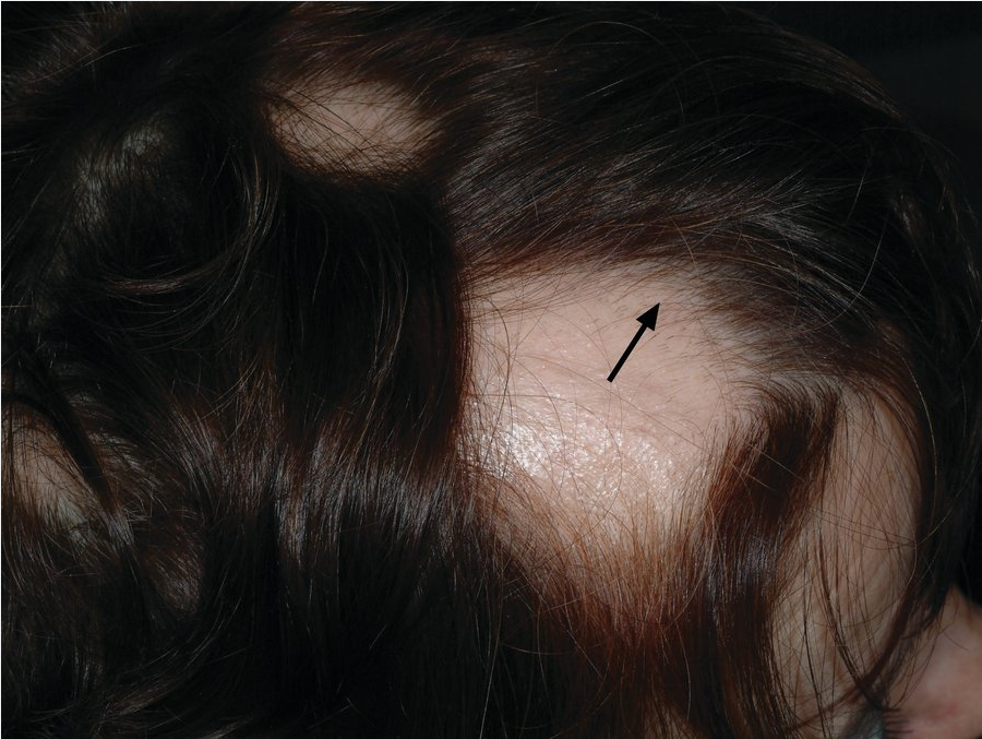 Well-demarcated, smooth, oval patches of hair loss. Note the smooth scalp and short hairs near the rim of the balding patch (arrow). Closer examination of these hairs will reveal “exclamation point” hairs (hairs that narrow close to the skin surface).
Traumatic alopecia includes traction alopecia, trichotillomania, and iatrogenic damage from chemicals and heat. Traumatic alopecia can begin as nonscarring; however, with continuing trauma, the alopecia can become permanent and scarring.
Androgenic alopecia, or patterned baldness, is due to the postpubertal terminal hair replacement, first with miniaturized follicles and eventually with atrophic follicles. It is seen in both men and women. Prevalence varies by populations studied. In comparison to white persons, pattern baldness is less common in Chinese, Japanese, and American Indians. The onset begins after puberty and can be gradual. In men, it presents as bitemporal hairline recession followed by vertex baldness (Figure 136). In women, it more often presents as generalized thinning rather than baldness and is often first recognized as widening of the part on the crown (Figure 137).
In properly selected patients, finasteride is used to treat androgenic alopecia. Dihydrotestosterone (DHT) binds to androgen receptors in hair follicles, transforming terminal hair follicles to miniaturized hair follicles. Finasteride inhibits the conversion of testosterone to DHT and slows the process of androgenic alopecia. In androgenic alopecia the pattern of hair loss reflects the sensitivity of the hair follicle to DHT.
Finasteride is contraindicated (FDA category X) in pregnant women because it is known to cause birth defects in the male fetus. Women who are or may potentially be pregnant should not take finasteride and should avoid contact with crushed or broken tablets because it can be absorbed through the skin.
Topical minoxidil can be used for male or female patterned hair loss. Minoxidil prolongs the duration of anagen, shortens telogen, and enlarges miniaturized follicles. Like finasteride, minoxidil is ineffective (and unnecessary) in hair loss caused by telogen effluvium.
Telogen effluvium is caused by a traumatic event (emotionally or physiologically) that disrupts the natural hair cycle resulting in diffuse hair shedding. The hairs on the scalp are all in different stages of the hair cycle, mostly anagen (growing) and telogen (resting). After a traumatic event, the hairs in the anagen phase are prematurely converted into the telogen phase. Hair shedding occurs about 3 months after the inciting event. This is most commonly seen in women in the postpartum period. In most cases, telogen effluvium is self-limiting with resumption of normal hair growth with resolution of the inciting factor. No specific therapy is required, and most cases will resolve spontaneously in about 6 to 12 months. Prognosis is especially good if the event causing the alopecia is identifiable.
Scarring Localized and Generalized Alopecia
Discoid lupus erythematous on the scalp can result in scarring localized alopecia. It presents as multiple pink, scaling hypo- and hyperpigmented scarring plaques (Figure 138) (see Common Rashes).
Acne keloidalis nuchae is a chronic inflammatory disorder of the follicles causing an alopecic scar on the nape of the neck (Figure 139). It can be pruritic. Pseudofolliculitis barbae occurs on the face and anterior neck. It is likely due to the curvature of the hair shaft and repeated trauma such as from shaving. When the curved hairs grow out after shaving, they curve downward into the skin, inciting an inflammatory response. This causes small pustules that can become keloidal. These conditions most commonly affect black men.
Lichen planopilaris is a diffuse scarring alopecia that more commonly affects postmenopausal women. It presents as mild scaling and limited erythema and scale surrounding the follicle (Figure 140). Associated symptoms are pain, burning, and pruritus.